

An easy guide to your eye’s anatomy
Your Eye's Surface
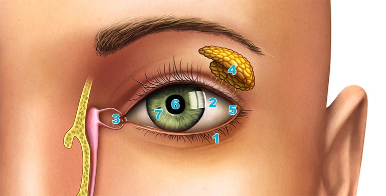
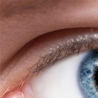
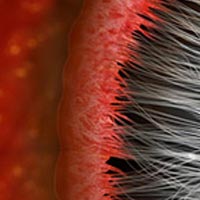
1. Eyelid
What is it?
Your eyelid is a thin fold of skin, lined with a row of eyelashes. Each eye has an upper and lower eyelid, and they can be opened and closed by special muscles.
What does it do?
The eyelids are vital to keeping your eyes healthy. By completely covering the front of your eye, they are able to block debris or impurities that may otherwise cause damage or infection.
Just as importantly, your eyelids help to prevent the surface of your eyes from drying out. They do this by regularly spreading tears when blinking, keeping the cornea moist.
2. Sclera
What is it?
You might better know the sclera by its more common name - the “white of the eye”. As its nickname suggests, this is the white part of your eye that surrounds the iris.

What does it do?
The sclera helps to support and protect the structure of your eye. It's made up of tough tissue, which ensures your eyeball keeps its shape.
Thanks to the sclera’s hardiness, the eyeball is less susceptible to injury than if it were surrounded by a softer tissue. It also provides support to the inside of your eye, by providing a surface for some of the ocular muscles to attach to.
3. Tear Duct
What is it?
The nasolacrimal duct is known to you and me as the tear duct. It’s the area found in the corner of your eyes, closest to the nose.

What does it do?
Your tear ducts carry excess tears away from the surface of your eyes. These tears are carried through to an area inside your nose called the nasolacrimal duct.
This explains why when you’re crying or suffering from watery eyes due to an allergy, you can sometimes taste the salt from your tears in your mouth. It’s also the reason why you might find your nose goes runny from crying.
4. Lacrimal Gland
What is it?
The lacrimal glands are shaped a bit like an almonds, found in the upper part of your eye sockets. You’ve got two lacrimal glands, one located just above each eye.
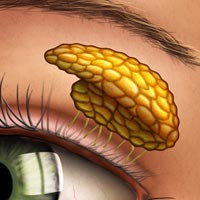
What does it do?
The basic function of your lacrimal glands is to produce tears. The secreted tears are collected on the conjunctiva of your upper eyelid.
Tears help to nourish and moisten your cornea (the outer layer of your eye’s surface). They also keep your eye clean, and lubricate it to avoid irritation.
5. Conjunctiva
What is it?
The conjunctiva makes up the lining inside your eyelids. It almost entirely covers your sclera, and is nourished by tiny blood vessels that are almost invisible to the naked eye.
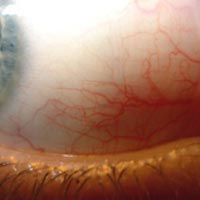
What does it do?
The conjunctiva acts as a vessel for your tears to be spread over the surface of your eye. This is important for ensuring your eyes are properly lubricated.
Conjunctivitis, also known as pinkeye, is a common condition associated with the conjunctiva. It occurs when the conjunctiva becomes inflamed, usually due to some sort of infection.
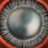
6. Pupil
What is it?
The pupil is the small black hole found in the centre of your eye. It is surrounded by the colourful iris, and leads directly to the inside of your eye.

What does it do?
The pupil acts as an entry point for light to enter your eye. Surrounding muscles in the iris, called the pupillae, adjust the pupil’s size automatically depending on light conditions.
In light conditions, your pupil will probably be around 3mm in diameter. In total darkness, this diameter can extend to over 6mm. This allows more light into your eye, allowing you to see more clearly in dimmer environments.
7. Iris
What is it?
The colourful ring found in the middle of your eye is called the iris. It’s made up of tiny pigment cells called melanin, which determine the colour of your eyes.
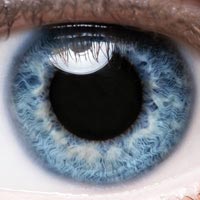
What does it do?
Your iris surrounds the pupil, and contains muscles that are able to alter its size. During light conditions, your iris causes the pupil to lessen in size. In darkness, the iris enlarges in order to allow more light to enter your eye.
The iris also acts as a wall that separates the anterior chamber (between cornea and iris) from the posterior chamber (between the iris and crystalline lens).
Inside Your Eye
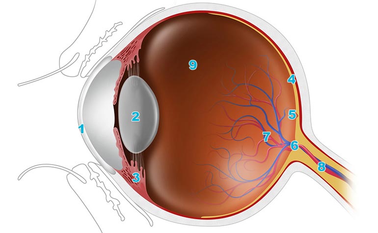
1. Cornea
What is it?
The cornea is a thin layer that covers the iris, pupil and anterior chamber. A healthy cornea is completely transparent, so that it can allow light through to the pupil.

What does it do?
Your cornea has a number of important roles. It is able to focus incoming light, making up around two-thirds of your eye’s total optical power.
Your cornea also protects the iris and pupil, preventing foreign objects from entering the inside of your eye. Additionally, it acts as a light filter, screening out many of the sun’s harmful rays and stopping them from reaching your retina where they’d otherwise cause damage.
2. Crystalline Lens
What is it?
The crystalline lens is a transparent structure that’s convex on both sides and suspended by tiny suspensory ligaments. It’s located just behind your iris, and is not visible from the outside without special viewing apparatus.
What does it do?
The surrounding ciliary muscles adjust the shape of the crystalline lens, allowing it to focus incoming light. This ensures that images are sharply focused on the retina at the back of your eye.
As the ciliary muscles contract, the suspensory ligaments that hold the lens in place relax. This causes the lens to assume a more spherical shape, allowing you to see close objects. When the muscles relax, the lens becomes flatter to allow you to focus on more distant objects.
3. Ciliary Body
What is it?
The ciliary body surrounds and is attached to the crystalline lens. Circular in shape, it separates the posterior chamber from the vitreous.
What does it do?
There are three main components that make up the ciliary body: the suspensory ligaments (zonules), ciliary muscle and ciliary processes. The ligaments and muscle work together in order to alter the shape of the crystalline lens and adjust how light is focused in your eye.
The ciliary processes produce aqueous humour. This is a transparent fluid that fills the anterior and posterior chambers of your eyes.
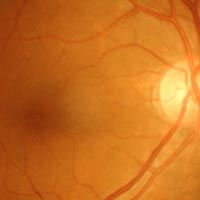
4. Retina
What is it?
Your retina is a light-sensitive layer that coats the inner part of your eye. It has a direct connection to your brain via the optic nerve.
What does it do?
Without the retina, your eye would have no way of interpreting the incoming light. When light hits the back of your eye, the retina processes and converts the energy into electrical signals.
The electrical signals are then sent to your brain through the optic nerve, ultimately translating into the images we see.
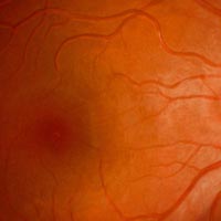
5. Macula
What is it?
Found on the surface of your retina, the macula is a sensitive, circular area that appears a bit darker than the space around it.
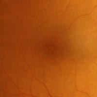
What does it do?
The macula contains a large amount of photoreceptors in comparison to the rest of retina. This means that it is suited to interpreting higher levels of and colour, providing the bulk of your central vision.
In a healthy eye, the macula is responsible for anything that requires the ability to see detail clearly. This might include reading or seeing the detail in someone's face.
6. Optic Disc
What is it?
The optic disc marks the beginning of the optic nerve as it enters the back of your eye. Only a few millimetres from the centre of your retina, a healthy optic disc is slightly oval in shape.
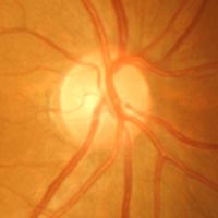
What does it do?
Because the optic disc is where the optic nerve has first contact with your retina, this is the point where the visual information begins its journey to the brain.
There aren’t any photoreceptors on the surface of your optic disc. Because of this, it is unable to process any visual information. This is the reason behind the ‘blind spot’ in your vision.
7. Central Retinal Vein and Artery
What are they?
The central retinal vein and artery line the back of the retinal wall. Healthy veins and arteries should appear consistent in width, with a deep red colour.
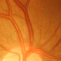
What do they do?
Your retina requires a blood supply in order to function. This is where the central retinal vein and artery comes in.
The central retinal artery supplies blood through the sclera, before branching out across the retina. Meanwhile, the central retinal vein carries blood and other waste products away from the retina. Both initially travel through the optic nerve in order to reach the retina.
8. Optic Nerve
What is it?
The optic nerve travels from your brain, all the way to the back of the eye. The point at which it meets your retinal wall is known as the optic disc.
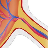
What does it do?
This is like the USB cable of your eye. After light has been converted into information by the retina’s photoreceptors, it is transmitted along the optic nerve.
It’s formed of around 1.2 million nerve fibres, that work together to send the impulses that will ultimately be processed into a visual image. The right side of your brain receives information from the left visual field of both eyes, while the left side receives information from the right visual field.
9. Vitreous
What is it?
Around 80% of your eyes’ total mass is made up of vitreous. This is a clear gel that fills the space between the crystalline lens and your retina.
What does it do?
The vitreous’ main function is to exert pressure on the inside of your eye. This helps to keep the layers of your retina tightly pressed together, as well as maintaining the general structure of the eyeball.
Your vitreous is completely stagnant, with no blood vessels to carry waste away. This means that anything that enters your eye will remain there unless surgically removed, possibly obscuring vision.
Now that you have a better understanding of how the eye works why not take a look at our vast range of eye care products. We have amazing products including eye drops, contact lens solutions & accessories that can help you maintain good eye health.