

Anatomy of the human eye
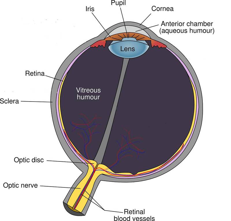
Parts of the human eye
The human eye is a sensory organ, sitting in the socket called the orbit, surrounded by skin, muscle and a strong layer of tissue. There are many parts of the eye that work together to make it work and, if looked after properly, protect it from some sight-threatening diseases and injuries.
Sclera
The sclera is the white part of the eye, a fibrous layer consisting of collagen that is continuous with the cornea.
Function of the sclera
The sclera surrounds most of the eye, front and back, and offers protection to the eye whilst conserving its structure and protecting it from injury.
Cornea
The cornea is the transparent dome shape that is the outer cover in front of the iris and pupil, behind a thin tear film.
Function of the cornea
The main function of the cornea is vision. As it is one of the first areas the light reaches when it enters the eyes, it is responsible for most of our eyesight. If the cornea is too flat, too curved or misshapen, it causes refractive errors, such as short or long-sightedness and astigmatism.
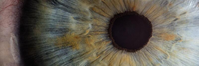
Iris
The iris is the brown, blue or green coloured part of the eye. It sits behind the cornea but in front of the lens and is circled around the opening known as pupil.
Function of the iris
The iris adjusts to different light settings, allowing the right amount of light through the pupil. It dilates in darker light conditions and constricts in brightly lit environments or when looking at close objects.

Lens
The crystalline lens is the transparent tissue that sits directly behind the pupil.
Function of the lens
When we are born, the lens is flexible and reshapes to allow the eyes to focus on varying distances. As we age, the lens loses its elasticity, which leads to the inevitable ageing condition presbyopia.
Vitreous humour
The vitreous humour is a gelatinous substance that lies behind the crystalline lens in the back of the eye. It contains a combination of water, collagen, and proteins.
Function of the vitreous humour
The vitreous humour has a vital role in ensuring the eye holds its spherical shape, thereby protecting your eye from injury and trauma. It takes up two thirds of the eye’s structure and attaches to the retina.
Retina
The retina is a light-sensitive tissue, lining the back of the eyes.
Function of the retina
This tissue works as a receptor for light. Like a film camera, the retina processes light through photoreceptor cells to detect colour and different lighting, which helps the optic nerve to send impulses to the brain, turning into images.
Optic nerve
The optic nerve is the biggest nerve of the eye. It sits at the very back of the eye and connects to the brain.
Function of the optic nerve
The optic nerve sends signals and impulses to the visual cortex of the brain, which is responsible for our vision. These impulses are transmitted through millions of fibres in the optical nerve.
How do the eyes work?
The various structures mentioned are all necessary to work together for us to perceive sight. If any of these parts are not working properly, due to eye conditions, diseases or injuries, our vision is affected.
Vision
The first step to eyesight is when light passes the thin tear film and through the cornea, where the eye first focuses. After the cornea, the light goes through the pupil (the opening of the iris), controlling how much light is let through.
The lens then works with the cornea to focus the light correctly, as it passes through the vitreous humour to the retina.
The retina forwards the received image through the optic nerve to the brain. Our brain then processes the received information and shows us the image – our eyesight.
Colour perception
Most people see the world in full colour, and they have their retina to thank for it.
The photoreceptors in the retina are equipped with two types of sensory cells, rods, and cones. The rods are responsible for processing changes in brightness and vision in low light environments, while variants of cones oversee processing colour vision.
The different variations of cones are each responsible for perceiving light that reacts to colour with varying wavelengths. Blue colours react with shorter wavelengths, green colours with medium wavelengths and red shades with long wavelengths.
The cones recognise each colour’s wavelengths and processes this information to the brain.
Parts perceptive to vision damaging conditions
From the front to the very back of the eye, every part is required to work together to offer us sharp vision. However, the eye is also a sensitive organ and, if not looked after properly, susceptible to many conditions.
Cornea
As the cornea is the most outer part of the eye, it is more susceptible to external dangers and surface diseases.
Injuries and abrasions can lead to corneal scarring and may cause distorted vision.
Allergies to pollen, pets or dust can irritate the outside of the eyes, causing them to water, itch or turn red.
The cornea can also be affected by dystrophies, such as keratoconus, a condition that causes the cornea to decrease in thickness and reshapes it, blurring and distorting your vision.
Retina
The retina lines the back of the eye and is therefore protected from most external factors, but it is still susceptible to damage.
Retinal diseases can be caused by various factors, such as age, trauma, general health conditions or genetics.
The most common retinal disorder is diabetic retinopathy, which is caused by the high blood sugar levels of type 1 or 2 diabetics.
Macular degeneration is another common eye condition that affects the macular (the centre of the retina). It is often age-related and affects the central field of vision.
