How Your Eyes Work
How Your Eyes Work
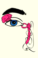
Interactive Tour of The Human Eye
How Your Eye Works
Welcome to our interactive tour of the human eye! Tap or click on one of the blue information points to begin your journey…
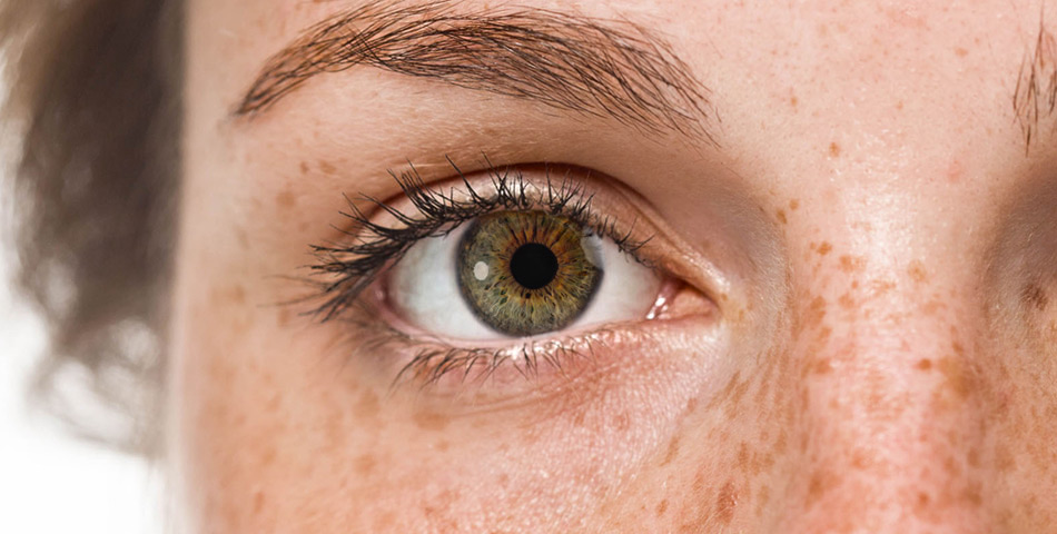
Eye Exterior
Eyelashes
Eyelashes
Eyelashes are hairs that help to block debris from entering your eye.
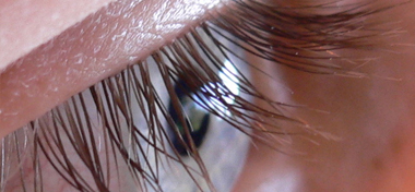
Eyelashes are hairs that help to block debris from entering your eye. They grow on the edge of the upper and lower eyelids. Both eyelids are lined with a row of eyelashes.
The lashes on the upper eyelid tend to be slightly longer than those on the lower lid. Growing up to around ten millimetres in length, the upper eyelashes usually curve upwards, while the lower eyelashes generally grow straighter.
The colour of eyelashes is often different to that of the head’s hair. However, they do tend to be darker for someone with dark hair, and lighter for someone with light hair.
How Do Eyelashes Block Debris?
To aid in blocking debris, eyelashes are sensitive to touch. When something touches the lashes, the eyelids will instinctively shut. In this way, they act as an early warning mechanism that allows the eyelids to quickly shut, before external debris can enter the eye and cause damage.
If an eyelash is ever removed, it can take over two months to regenerate. The eyelash follicles are associated with sweat glands, which can cause styes when blocked or infected.
Eyelid
Eyelid
The eyelids block external debris and help to keep your eye lubricated.
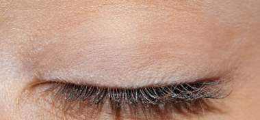
The eyelids block external debris and help to lubricate your eye.
They are capable of opening and closing both voluntarily and involuntarily.
The blink reflex acts as a semi-autonomous way to block foreign bodies entering the eye, assisted by the eyelashes.
The eyelid regularly spreads tears and other fluids across your eye’s surface. This keeps your eye lubricated, and ensures the cornea is given the required nutrients to function correctly. Eyelids are also vital to prevent the eyes from drying out during sleep.
Structure of the Eyelids
The eyelid consists of an anterior and posterior portion. The anterior portion contains the eyelashes and glands, while the posterior portion contains the tarsal conjunctiva and tarsal plate. The tarsal plate is composed of dense, fibrous tissue, which maintains the structural integrity of the eyelid.
The surface skin of the eyelid is much thinner than most of that across the rest of the body. It contains high concentrations of sebaceous glands. These sebaceous glands secrete an oily substance called sebum, which helps to lubricate the skin - something especially important in the eyelids.
The eyelid contains three types of secretary glands. The glands of Moll are sweat glands, while the meibomian glands and glands of Zeis are oil-producing glands. These oil-producing glands act to lubricate and waterproof the skin, as well as providing protection against bacteria.
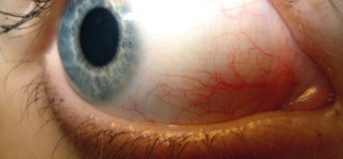
Sclera
Sclera
The sclera protects and supports the structure of your eye.
Commonly known as the “white of the eye,” the sclera supports and protects the structure of your eye. The sclera makes up most of the outer layer of the eyeball and optic nerve.
The tough tissue of the sclera maintains the shape of the eyeball. It also helps to protect against injury, and provides an attachment for 6 of the eye’s extra ocular muscles. It is covered by the clear conjunctiva, and is continuous with the cornea.
The sclera is supplied with nutrition from the underlying choroid and overlying episclera (a thin layer of tissue that covers the sclera). It is also supplied with a high density of nerves - damage to these nerves can cause a significant amount of pain.
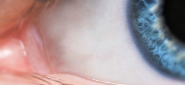
Tear Duct
Tear Duct
The tear duct drains excess tears into the nasal cavity.
The tear duct (also known as puncta) are small openings that drain tears into the nasal cavity.
Tears are released on the surface of the eye by the lacrimal glands. These tears help to clean and lubricate the eye, while also nourishing and moistening the cornea.
When the eyelids blink, the lids push the tears across the eye and into the puncta. Draining these excess tears is important for regulating moisture levels in the eye.
The Journey of Tears
The tears then travel through small canals in the lids to a sac attached to the side of the nose (lacrimal sac), then down a duct (the nasolacrimal duct) before emptying into your nose, where they evaporate or are reabsorbed.
Strong emotion, as seen in crying, can cause tears to be produced at a faster rate than they can be drained.
A blocked tear duct is a common condition that can occur when the duct is obstructed in some way. This can result in tears being unable to drain properly, which causes the eye to become watery, irritated, and occasionally infected.
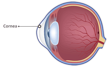
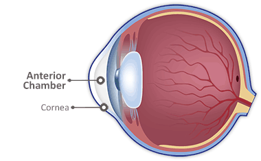
Cornea
Cornea
Cornea
The cornea protects the iris and pupil, while providing much of the eye’s focusing power.
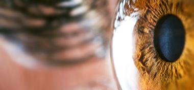
The cornea protects the iris and pupil, while providing much of the eye’s focusing power. Dome-shaped and clear, the cornea covers the pupil, iris and anterior chamber.
The cornea is transparent in order to refract light correctly. The presence of even the smallest blood vessels would interfere with this. Any cloudiness or opaqueness can heavily interfere with vision.
The cornea also serves as a filter, screening out damaging ultraviolet (UV) wavelengths in sunlight. Without this protection, the lens and retina would be susceptible to injury from these UV rays.
Nourishment of the Cornea
The cornea has many layers of proteins and cells. It has no means of blood supply, meaning it must be supported by other sources.
The aqueous humor is one source of nourishment for the cornea, bringing metabolic supplies such as glucose. It flows through the leaky barrier presented by the endothelium and enters the stroma, before delivering its contents to the cells there.
The cornea is also nourished by oxygen. The oxygen supply comes from two directions; in front where the tear film is in contact with the outside environment (or the conjunctiva during sleep), and from the aqueous behind.
Anatomy of the Cornea
The five layers of the cornea consist of:
- Epithelium - The outermost layer, which helps to prevent impurities from entering the eye. The epitheliumIt is made up of five or six layers of cells that are bound together tight enough to be waterproof.
Though the epithelium only constitutes around 10% of cornea’s volume, it consumes 40% of the cornea’s total oxygen requirement. Scarring cannot occur on this layer, as the epithelial cells regenerate with an average life of one week. - Bowman’s Layer - Made up of protein fibers, the Bowman’s layer is tough, helping to shape the rest of the cornea. Made up of strong, layered protein fibers called collagen, this layer is able to scar through injury.
- Stroma - By far the thickest layer of the cornea (90% of the corneal thickness), the stroma consists of 78% water. The stroma also contains keratocytes, which help to repair the cornea if it gets damaged.
Collagen makes up 16% of the stroma, helping to give the cornea its elasticity and form. The unique shape, arrangement and spacing of the collagen are essential in producing the cornea’s transparency. - Descemet’s Membrane - This membrane acts as another protective layer against impurities and injury.
- Endothelium - The innermost layer, the endothelium regulates the fluid in the cornea. Pumping excess fluid out of the stroma, the endothelium ensures that the stroma remains clear and haze-free.
While the cornea contains no blood vessels, it is filled with sensory nerve fibres. Pain receptors in the cornea have been suggested to be 300-600 times more dense than skin, making injury to the cornea extremely painful.
Conjunctiva
Conjunctiva
The conjunctiva produces mucins and tears to keep the eye lubricated.
The conjunctiva lines the inside of the upper and lower eyelids, producing mucins and tears to keep the eye lubricated. Continuous with the cornea, the conjunctiva lies on top of the sclera.
Mucins produced by the conjunctiva maintain a healthy tear film. The conjunctiva plays a vital role in the defence of the outside eye, as mucin secreted by the conjunctiva helps to trap and clear allergens.
Nourished by tiny blood vessels (almost invisible to the naked eye), the conjunctiva is transparent. The tears and mucins released from the conjunctiva reduce the friction from blinking, while nourishing the eye.
Anatomy of the Conjunctiva
There are three main parts that make up the anatomy of the conjunctiva:
- Palpebral conjunctiva - The lining on the underside of your eyelids.
- Bulbar conjunctiva - The covering of the sclera, that moves with your eye’s movements.
- Fornix conjunctiva - The loose folds that connect the bulbar conjunctiva to the palpebral conjunctiva to form a sort of sac. The fornix conjunctiva is very flexible, allowing the eyelids and eyeball to move freely.
Conjunctivitis, also known as pinkeye, is a common condition caused by an infection or allergic reaction. The conjunctiva becomes inflamed, often causing the eye to appear pink or red. Symptoms of pinkeye can also include watering and swelling.
Meibomian Glands
the Meibomian Glands
The meibomian glands produce an oily substance that prevents evaporation of the tear film.
The meibomian glands, found at the rim of the eyelids, are a type of sebaceous gland that produces meibum. This oily substance helps to prevent evaporation of the tear film.
A type of sebaceous gland (glands that secrete an oil), the meibomian glands produce an oily substance called meibum.
Without meibum, the eyes would dry out. The substance also traps tears between the edge of the eyelid and the eye, helping to spread nutrients to the cornea.
Meibum and Sleep
During sleep, the eyes aren’t lubricated by blinking. Therefore, it’s important for the existing fluids to remain in the eye. The meibum ensures your eyelids are airtight when closed, which helps keep moisture in.
Dry eye syndrome is a common eye condition, and can be caused by a dysfunction in the meibomian glands. If the glands become inflamed, they can become blocked and reduce their effectiveness.
Iris
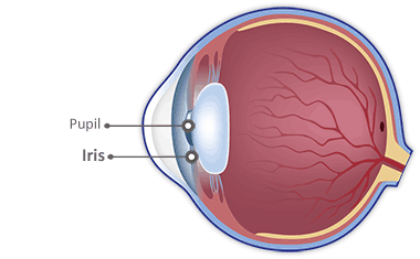
Pupil
Pupil
The pupil is the small black hole visible at the centre of the iris, and controls how much light enters your eye.
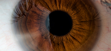
Located in the centre of the iris, the pupil is a small hole that controls how much light enters your eye. Unlike the colours of the iris, the pupil appears totally black.
The pupil changes size in order to control how much light hits the retina at the back of the eye. It is controlled by two groups of surrounding muscles, known as the sphincter pupillae and the dilator pupillae.
The sphincter pupillae contract in bright light conditions, decreasing the size of the pupil, while the dilator pupillae cause the pupil to grow larger in dim light conditions when they contract.
Generally, the pupil’s diameter will be around 3 millimetres in light conditions, and around 6 millimetres in the dark. However, the size of the pupil can greatly vary depending on the individual and their age.
Sending Signals To The Pupil
Light-sensitive cells in the retina react to light hitting them and send signals along the oculomotor nerve to the circular sphincter pupillae to reduce the size of the pupil. This automated process is known as the pupillary light reflex.
The sympathetic pathway controls the dilator pupillae, beginning in the brain. Information is passed from here via the spinal cord and back up to the dilator pupillae, in order to dilate the pupil.
The autonomic nervous system (a control system that we use unconsciously) regulates many of the pupil’s actions, and is responsible for the involuntary functions of the human body. It can be divided into two divisions, which are as follows:
- The parasympathetic nervous system (PN) is responsible for the body’s “rest and digest” function.
- The sympathetic nervous system (SNS) is responsible for the body’s “fight or flight” response.
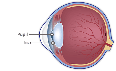
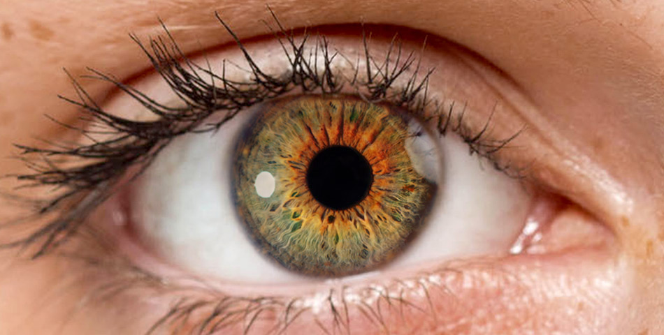
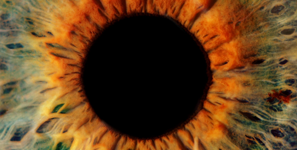
Iris
Iris
The iris is the coloured section at the front of the eye, which controls the size of the pupil.
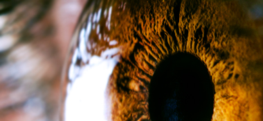
The iris controls the size of the pupil. By changing the size and diameter of the pupil, the iris adjusts the amount of light that enters your eye.
The iris surrounds the pupil, and is attached to the middle of the anterior surface of ciliary body. It separates the anterior chamber (between the cornea and iris) from the posterior chamber (between the iris and lens).
What Is the Iris Made Of?
The colour of the iris comes from microscopic pigment cells called melanin. The texture of your iris is as unique as your fingerprints.
There are four layers that make up the iris; the anterior limiting layer, the stroma, the anterior pigmented epithelium and the posterior pigmented epithelium.
The stroma forms the main bulk of the iris and contains the sphincter pupillae, dilator pupillae muscles, vessels and nerves.
The sphincter muscle lies around the edge of the pupil. When light conditions brighten, the sphincter contracts, causing the pupil to constrict. The dilator muscle runs radially through the iris, similarly to spokes on a wheel. This muscle causes the pupil to dilate in dim lighting.
The pigment epithelial layer lies at the back of the iris. This layer is just two cells thick, and also contains pigment granules, except in albino eyes. The epithelial layer blocks light from entering the eye through anything but the pupil.
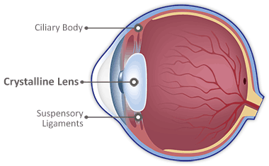
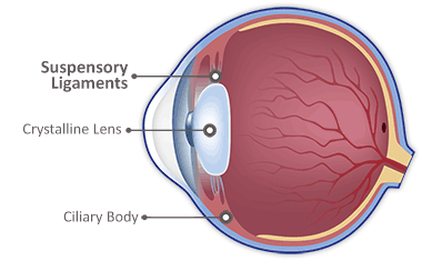
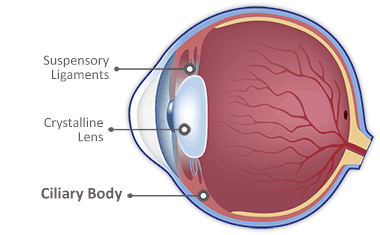
Crystalline Lens
Crystalline Lens
Crystalline Lens
The crystalline lens focuses the light that enters your eye onto the back of your eye.
The crystalline lens focuses the light that enters your eye. The lens itself is a transparent, biconvex (convex on both sides) structure located behind the iris. Ciliary muscles change the shape of the crystalline lens, focussing light so that images come to a sharp focus on the retina.
The shape of the crystalline lens is controlled by the ciliary muscle, which is attached to the suspensory ligaments (also called the zonules of Zinn) that hold the lens in place. These are attached at one end to the lens capsule, with the other attached to the surrounding ciliary muscle.
When the ciliary muscles relax, the zonules become taut and the crystalline lens is stretched to form a flattened shape with a weaker focusing power. As the ciliary muscles contract, the zonules loosen and the lens assumes a more spherical shape.
Anatomy of the Crystalline Lens
The crystalline lens has a diameter of around 10mm, and a thickness of around 4mm. The lens itself is enclosed by the lens capsule. Made of collagen, the lens capsule is elastic, allowing the crystalline lens to be shaped by the suspensory ligaments.
The layer just past the lens capsule is the lens epithelium. This regulates the liquids and nutrients in the crystalline lens, so that its internal conditions remain constant and stable. It also has the task of dividing in order to create new lens fibres.
Most of the crystalline lens is made up of lens fibres. Densely packed, these fibres are transparent to allow light to pass through. The fibres are arranged in concentric layers, much like the layers of an onion.
Suspensory Ligaments
Suspensory Ligaments
The suspensory ligaments connect the ciliary muscles to the crystalline lens, holding it in place and helping to change its size.
The suspensory ligaments (also called “zonules”) connect the ciliary muscles to the crystalline lens. The zonules are very strong, holding the lens in place and helping to adjust its size.
The zonules and ciliary muscles are able to work together to adjust the shape of the crystalline lens. The curvature of the lens is fine-tuned by them, increasing or decreasing the refraction of light to focus at different distances.
Zonules and the Ciliary Muscle
The ciliary muscle relaxes when your eye looks at a distant object, which pulls on the suspensory ligaments. This in turn pulls on the crystalline lens, causing it to become flatter and more able to focus light from a distant object.
The ciliary muscle contracts when the eye looks at a close object. This releases tension from the suspensory ligaments. The crystalline lens becomes more convex (rounded/curved), allowing it to focus light from the close object onto the retina.
This adjustment in lens shape, to focus at various distances, is known as accommodation.
Anterior Chamber
Anterior Chamber
The anterior chamber contains a fluid called aqueous humor, which provides nourishment to the cornea and lens.
The anterior chamber acts as a channel for the aqueous humor to travel through, which provides nourishment to the cornea and lens. The aqueous humour also removes waste products from the front of the eye.
The chamber is located between the iris and the inner layer of the cornea.
What is the Aqueous Humor?
Made up of 98% water, the aqueous humor contains electrolytes, ascorbic acid and amino acids. The fluid provides nutrients and oxygen to any structures without their own blood supply. It also helps to maintain pressure and keep the anterior and posterior chamber inflated.
In order to maintain pressure, the aqueous humour has to be drained at the same rate as it’s produced. After entering the anterior chamber, the aqueous humour exits into “Schlemm’s canal”, a channel found where the sclera and cornea join together.
Ciliary Body
Ciliary Body
The Ciliary Body controls the shape of the crystalline lens and produces a fluid called the aqueous humor.
The ciliary body controls the shape of the crystalline lens and produces a fluid called the aqueous humor. Circular in shape, the ciliary body separates the posterior chamber from the vitreous. It is made up of:
- The Ciliary Muscle
- Zonular Fibres
- Ciliary Processes
The ciliary muscle is attached to the crystalline lens via the zonular fibres. Together, they control the shape of the lens, causing it to contract or relax when viewing either a distant or near object. By shaping the lens, it focuses light onto the back of the eye.
The ciliary processes produce aqueous humour, which provides nourishment for the lens and cornea.
Flow Of The Aqueous Humor
The aqueous humor flows from the ciliary body through the pupil and into the anterior chamber. It diffuses into the trabecular meshwork and into the canal of schlemm, then finally into the tiny blood vessels surrounding the eye.
Eye pressure depends on the production and re-absorption of aqueous humor. As the ciliary body is responsible for this, it is often the target structure for medications.
The ciliary body is part of the uvea, which is also known as the middle layer of tissue surrounding the eye. The uvea consists of the iris, ciliary body and choroid, and is largely responsible for providing nutrition and oxygen to other parts of the eye.
Retina
Vitreous
Vitreous
The vitreous is a clear gel that exerts enough pressure to keep the retinal layers tightly pressed together.
The vitreous (also referred to as the vitreous humour or vitreous body) is a clear gel that exerts pressure on the inside of your eye. The main function of this pressure is to keep the retinal layers tightly pressed together.
The pressure also helps to maintain the round shape of the eye. It is the largest structure within the eye, taking up around 80% of the eye’s total volume. The vitreous is transparent, allowing light to pass through it onto the retina.
What is the Vitreous Made Of?
The vitreous contains no blood vessels, and 99% of its volume is water. The remaining 1% is made up of collagen, organic salts and lipids (a group of naturally occurring molecules). The collagen gives the vitreous its jelly-like properties.
The structure of the vitreous changes as the eye ages - the vitreous of a child’s eye is more solid than that of an older eye. As your eyes age, collagen within the vitreous shrinks, resulting in areas of dense gel that move with eye movement.
This casts a shadow on the retina and results in the spidery floaters that most of us notice in bright light conditions.
The vitreous is stagnant, meaning that anything that enters the gel will remain there unless surgically removed. 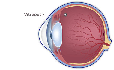
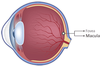
Fovea
Fovea
The fovea is a small part of the macula that provides the sharpest and most accurate colour vision of the retina.
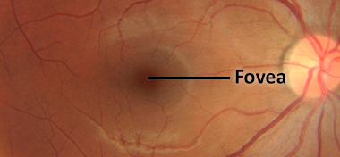
The fovea is the part of the retina that provides sharp and accurate colour vision. It makes up less than 1% of the retina, but information received here takes up over 50% of the Visual Cortex in the brain.
Where is the Fovea?
The fovea is the small dip in the retina noted at the centre of the macula roughly 1.5mm in size and circular in shape. When a person looks directly at an object, its image is focused on the foveola (found within the fovea), helping to produce a clear, detailed image.
The Fovea contains the largest amount of cone photoreceptors (which detect colour) in the retina and is also part of the retinal avascular (no blood vessels) zone. This allows light to pass through to the deeper layers of the retina without any dispersion or loss.
The Foveola
The foveola is the thinnest part of the retina and is found at the centre of the fovea. Only 0.35mm in size, it is the only area of the retina with enough cones to provide sharp, accurate colour vision.
The foveola is a rod- free zone and exclusively contains cone photoreceptors, with a lower density of blue cones. The cones in this area are packed extremely close together compared to anywhere else in the retina, to achieve maximum visual acuity.
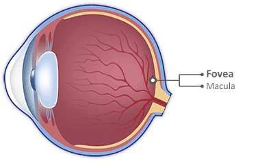
Macula
Macula
The Macula produces your central vision, providing detail and colour.
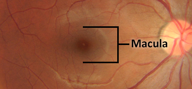
The Macula is a highly sensitive circular area that is responsible for providing detail and colour to your vision in bright light conditions. The macula is able to provide such detailed vision because of its high concentration of photoreceptors, particularly cone cells, found at its centre.
The Macula is the part of the retina that produces your central vision. For most people, it is the structure of the eye that is used to detect detail and for reading/close work.
Inside the Macula
The macula area is made up of the fovea, foveola, foveal avascular zone (FAZ), and the umbo. These structures are directly in the line of sight. When a person looks directly at an object, its image is focused on the foveola.
Natural antioxidants (lutein and zeaxanthin) are found within the macula, helping to absorb damaging, high-energy blue and ultraviolet light that can potentially destroy cells and play a role in many diseases.
Conditions Of The Macula
Damage to the macula can result in loss of central vision, which is usually immediately obvious and can be described as either an obstruction to the central vision/blurred central patch or metamorphopsia. This describes a distortion of images; straight lines appear wavy/jagged or pulled out of position.
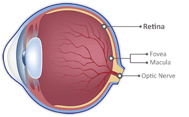
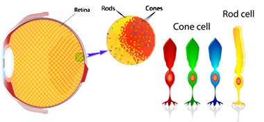
Retina
Retina
The retina converts light into signals that are passed on to the brain.
Cells within the retina process and convert light energy into electrical signals. These electrical signals are then transmitted to the brain through the optic nerve and interpreted as the images we see.
Visible structures that you will find on the retina include:
- Optic Disc
- The Macula
- The Fovea
- Central Retinal Artery and its branches
- Central Retinal Vein and its branches
The Layers of the Retina
The retina is composed of ten layers. The only layer that is directly sensitive to light is the photoreceptor layer. The other nine layers are transparent so that light entering the eye can pass directly through to the deeper photoreceptor layer (rods and cones), where the conversion of light energy begins.
The retinal layers from the outside (nearest the blood vessel enriched choroid) to the inside (nearest the vitreous humor) are named as such:
- Pigmented epithelium - These cells contain melanin, which absorb light and decrease light scatter within the eye.
- Photoreceptors (Rods and Cones).
- External (outer) limiting membrane.
- Outer nuclear layer.
- Outer plexiform layer.
- Inner nuclear layer.
- Inner plexiform layer.
- Ganglion cell layer.
- Nerve fiber layer.
- Internal limiting membrane (the boundary between the retina and the vitreous).
Light is converted into neural signals by the photoreceptor layer. This energy is transmitted to the middle layers of the retina containing bipolar cells. The signal is then relayed to the inner layers of the retina, consisting of multiple different types of ganglion cells (the cells that transmit to the brain).
The axons of the ganglion cells form the optic nerve and carry all of this information through the to the brain.
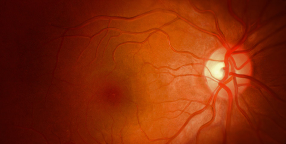
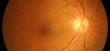
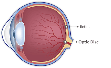
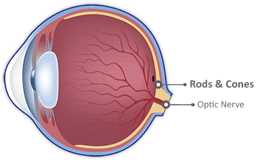
Optic Disc
Optic Disc
Optic Disc
The optic disc marks the entrance of the optic nerve into the retina.
The optic disc (also known as the optic nerve head or blind spot) is the beginning and innermost section of the optic nerve in the retina.
Typically, the optic disc is vertically oval in shape, and can be found 3 to 4 millimetres away from the center of the retina.
The “Blind Spot”
There are no photoreceptors (rods and cones) at the optic disc, which means that this area of the retina doesn’t respond to light. As a result, the optic disc is known as your “blind spot”.
Retinal nerve fibres are spread across the surface of the retina in a thin layer known as the nerve fibre layer. The nerve fibres come together on the edge of the optic disc disc and then move down its inner surface. This compressed area of nerve fibres is seen as the neuroretinal rim.
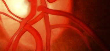
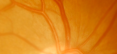
The disc itself has an orange-pink coloured rim known as the neuroretinal rim. Visual impulses are carried through here and sent to the brain to be processed. Major blood vessels that supply the retina (central retinal artery and central retinal vein) also exit the retina at this point.
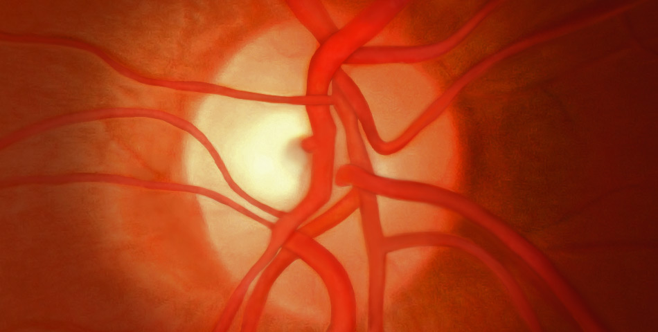
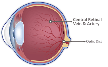
Central Retinal Vein and Artery
Central Retinal Vein and Artery
The central retinal vein and artery provide a blood supply for the retina.
The central retinal vein and artery provide a blood supply for the retina. The retina requires a constant blood supply, and this makes sure that the cells of the retina get all the nutrients they need to continue working.
The retina is supplied by the central retinal artery and the posterior ciliary arteries. These are all branches of the ophthalmic artery, which supplies all structures found within the eye socket.
- The central retinal vein’s function is to carry blood and waste products away from the retina, while the central retinal artery carries blood to the retina.
- The central retinal artery travels in the optic nerve through the sclera and then branches out to supply the layers of the inner retina.
The outer and middle retinal layers, are nourished by branches of the posterior ciliary arteries. These enter the back of the eye, outside the optic nerve. These vessels also supply the choroidal layer, which is behind the retina.
The central retinal arteries are narrower and brighter than the veins. After entering through the optic disc, the arteries send branches across the surface of the retina.
Artery Or Arteriole?
Unlike true arteries in the body, the central retinal artery lacks a muscular coat. Therefore, “arteriole” is a more accurate term for the retinal artery.
Like the retinal artery, the central retinal vein also enters the retina through the optic disc. The central retinal vein functions to drain blood from the capillaries of the retina into the ophthalmic vein.
The central retinal vein and artery are commonly associated with central retinal vein occlusion (CRVO) or central retinal artery occlusion (CRAO). If either suffers from a blockage, the retina may undergo severe damage and produce conditions that can threaten eyesight.
Rods and Cones
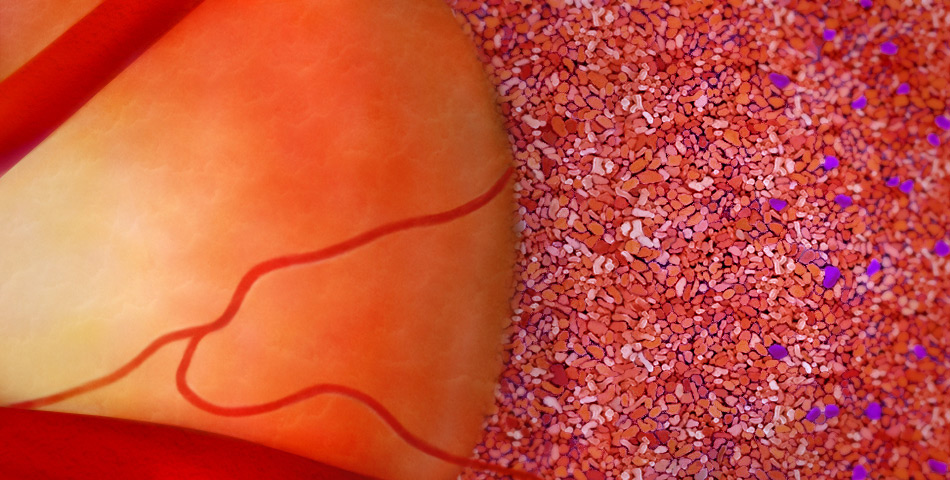
Rods and Cones
Rods and Cones
Rods and cones convert light into nerve signals that are interpreted as visual images by your brain.
Rods and cones are collectively known as photoreceptors. Photoreceptors convert light into nerve signals that are then delivered to the brain. The brain is able to process the signals into the images that you see.
Most people have roughly 7 million cones and 120 million rods in each eye.
What’s The Difference Between Rods And Cones?
Cones are found mainly within the centre of the fovea and are responsible for your colour vision in bright light conditions, helping you to see colour and detail during the day.
There are three types of cones that can be found within the retina, each with a photopigment that is sensitive to either a red (Long), green (Medium) or blue (Short) wavelength of light. These three types of cones work together to help you perceive a full spectrum of colour.
Rods are found in the periphery of the retina. They have a greater sensitivity to dim light, helping you to see in the dark. Rods are not sensitive to colour but are over 1,000 times more light-sensitive than cones.
At night and in low light conditions your sense of vision comes from the rods alone, which is why you will find it difficult to see colour at night.
Damaged cones can result in a colour deficiency. Damaged rods can cause problems seeing in the dark and at night.
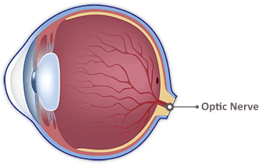
Optic Nerve
Optic Nerve
The optic nerve transmits impulses received from the retina to the visual cortex in the brain, forming an image in our minds.
The optic nerve transmits impulses received from the retina to the visual cortex in the brain, forming an image in our minds.
Light is converted into impulses via photoreceptors found within the retina. This information is sent through the optic nerve and past the optic chiasm.
The optic nerve at this point becomes the optic tract; from here, impulses are sent to the brain to be processed as a visual image.
Visual Information
The optic nerve is formed of 1.2 million nerve fibres that begin in the nerve fibre layer of the retina. The retinal nerve fiber layer is the 9th layer of the retina (closest to the vitreous).
Nerve signals travel along the optic nerve from each eye. The two optic nerves meet at the optic chiasm - here the optic nerves from each eye divide. Half of the nerve fibers from each side cross to the other side.
Because of this arrangement, the right side of the brain receives information from the left visual field of both eyes, and the left side of the brain receives information from the right visual field of both eyes.
Damage to the optic nerve can cause permanent and potentially severe loss of vision. The type of visual field loss will depend on which parts of the optic nerve were damaged.
Optic Nerve

Optic Nerve
Optic Nerve
The optic nerve transmits impulses received from the retina to the visual cortex in the brain, forming an image in our minds.
The optic nerve transmits impulses received from the retina to the visual cortex in the brain, forming an image in our minds.
Light is converted into impulses via photoreceptors found within the retina. This information is sent through the optic nerve and past the optic chiasm.
The optic nerve at this point becomes the optic tract; from here, impulses are sent to the brain to be processed as a visual image.
Visual Information
The optic nerve is formed of 1.2 million nerve fibres that begin in the nerve fibre layer of the retina. The retinal nerve fiber layer is the 9th layer of the retina (closest to the vitreous).
Nerve signals travel along the optic nerve from each eye. The two optic nerves meet at the optic chiasm - here the optic nerves from each eye divide. Half of the nerve fibers from each side cross to the other side.
Because of this arrangement, the right side of the brain receives information from the left visual field of both eyes, and the left side of the brain receives information from the right visual field of both eyes.
Damage to the optic nerve can cause permanent and potentially severe loss of vision. The type of visual field loss will depend on which parts of the optic nerve were damaged.
Reversed Image
Reversed Image
Images transmitted from the retina are upside-down and reversed, meaning your brain must reorient them.
Images transmitted from the retina are upside-down and reversed. This means that the brain must interpret the image, reorienting it correctly.
Having the image reversed and upside-down on the retina is actually an evolutionary advantage. The crossing of light rays diverging from different points on an object causes the image of the visual field to be inverted. The visual field is also left-right reversed on the retinal surface as the rays are focused.
Why Are The Images Reversed?
The diagram below illustrates how we are able to see large objects, and why the resulting image must be reversed:

From the inside of the eye, the top of the building is projected on to the lower part of the retina, while the bottom of the building is projected on the upper part of the retina.
Rather than trying to coordinate with an upside-down, reversed world, the brain processes the visual information to orient everything correctly. The brain is used to seeing the upside-down image, and is able to compensate.
Because of this, it’s believed that babies see the world upside down for the first few days after being born. Their brains are still getting used to the concept of vision, and therefore have not adapted to the reversed image.
The Mind’s Eye

The Mind’s Eye
The Mind’s Eye
This is the final image that your brain interprets from the signals sent from your eyes.
The retina sends an image to the brain that is reversed and upside-down. Because of this, the brain must interpret these signals and reorient images correctly.
Signals from the retina are sent along the optic nerve. The two optic nerves travel from both of your eyes to meet at the optic chiasm. The optic nerves from each eye then divide, with half of the nerve fibres from each side crossing to the other side.
This means that the right side of the brain receives information from the left visual field of both eyes. The left side of the brain receives information from the right visual field of both eyes.
Inside the Brain
The vast majority of nerve fibres in the optic tract project to the lateral geniculate nucleus (LGN). The LGN relays information to the primary visual cortex (located at the back of the brain’s occipital lobe).
The primary visual cortex sends a large amount of information to the secondary visual cortex. The primary and secondary visual cortex are responsible for reconstructing the image received from the retina.
The visual analysis that begins in the primary and secondary cortex passes through two major systems for processing. The first is the ventral pathway which is thought to be involved in recognizing objects. The second is the dorsal pathway, which appears to be essential for locating objects.
Contents
![]()
An interactive tour of the human eye is brought to you by Lenstore. Our eyes are among the most complex organs in our bodies. Full of parts with long names and obscure functions, it can be difficult to grasp exactly how our eyes are able to see.
In our ongoing mission to educate the world about vision and eye care, we’ve created our very own interactive eye. Highly visual and interactive, this educational resource lets you explore the human eye and learn about one of our most important senses.
If you are a contact lenses wearer then please have a look through our range of branded contact lenses available at a huge discount to the high street.